little time left. The folks at Womens' International Pharmacy have made it easy by posting links on their website. Go to http://www.womensinternational.com and just follow the instructions.


Here's a sample of some of the objections citizens have raised to Wyeth's petition:


I have been taking bioidentical hormones for two years. Bioidentical Hormones have been a great relief to me without the risk. I consult with my Physician who prescribes bio-identical hormones specifically for me, and my pharmacist prepares them. Without this medication and I would not be able to sleep; I would not be able to work due to the constant hot flashes. Without this medication, I find that I have less tolerance and I am considerably disagreeable. I also have problem with my memory without them. I want the bioidentcial hormones for the health benefits they provide. I urge you to not be swayed by Wyeth's petition. The product Premarin made by Wyeth, is made from pregnant horses not natural sources. Wyeth's hormones have been shown to cause cancer. I would not expect my government and its officials to submit to the highly funded petitioning of a pharmaceutical company who product is threatened by bioidentcial hormones. I do not expect my government to approved Wyeth's petition and leave me no choice of bioidentcial hormones and only the choice of Wyeth's cancer causing drugs Preamrin and Prempro. I ask that the FDA reject Wyeth's petition Docket #2005P-0411.

Another petitioner writes:

As a woman I take exception to Wyeth accusing the Compounding Pharmacy industry of unsafe practices. As a citizen of the United States I expect the FDA to stand up for my rights and the rights of all women who have found or in the future may seek consistent, safe and effective treatment with bioidentical hormones. Eliminating options by bowing to a large pharmaceutical company like Wyeth is not in the public interest and would deprive hundreds of thousands of American women from access to bioidentical hormones. Synthetic hormone replacement has been proven unequivocally unsafe in a government sponsored study and should not be forced as the sole treatment option for women. I hereby request the FDA rule against Wyeth's request. The FDA should not close down the bioidentical option of healthcare. I welcome studies of bioidentical hormones even though they are already FDA-approved and have been working effectively for decades. We already have the proof - hundreds of thousands of women, who over the past two decades have chosen bioidentical hormones based on their physicians' assessments. They are living proof that bioidentical hormones are safer and more effective and reliable than synthetic hormone drugs.

A physician and user of bio-identical hormones writes:

Wyeth, the filer of this complaint, is trying to prevent women from being able to choose less expensive compounded options for hormone replacement. There is medical evidence that in modifying the structure of their drugs (such as Premarin and Prempro) so that they could be patented, they may have introduced factors that cause the health risks identified in the Women's Health Initiative. This complaint appears to be filed for commercial purposes because of the market share that has shifted from Wyeth's products to bio-identical products from compounding pharmacies. If the complaint were upheld, patients and their doctors would not have a choice in hormone treatments. Wythe's commercial strategy of trying to eliminate the 'competition' from compounding pharmacies is against the public interest and in the interest of its own corporate profits. Women and their doctors should be able to choose between patented formulations such as those offered by Wyeth, bioidentical formulas available from compounding pharmacies, and no hormone treatment. I have been taking bio-identical hormones for several years and have had excellent results in improving my symptoms. I have been unable to take other synthetic hormones in the past, and am very concerned that my best treatment option will be taken away.

If you get a 64-slice CT coronary angiogram

With new 64-slice CT scanners popping up everywhere nowadays, be sure to get your heart scan with it.

The new scanners do indeed provide wonderful images of the coronary arteries. But, say you have a 20% blockage in one artery by a coronary angiogram generated on one of these devices. What will you do in 1, 2, or 3 years when you want to know if you have progressed? Should you have the CT angiogram repeated?

Well, if you did you'll be exposed to a large dose of radiation--appropriate for a diagnostic test, but not for a screening test. The radiation exposure is not that different from undergoing a full conventional cardiac catheterization, or up to 100 chest x-rays.

"20% blockage" is also, contrary to popular opinion, not a quantitative measure. It is just an estimate of the diameter reduction at one spot. That number says nothing about the lengthwise extent of plaque. It also says nothing about the potential for "remodeling", the phenomenon of artery enlargement that occurs as plaque grows. In other words, if you had another CT coronary angiogram a year later and was told that your blockag was still 20%, in reality you could have had substantial plaque growth but it would not be reflected in that value.

People will come to me after having a CT angiogram for an opinion. Unfortunately, I send them back to their scan center to get a simple coronary calcium score. That measure is easy, quantitative, precise, and can be repeated yearly if necessary to track progression. (Track Your Plaque--I hope most of you get this by now.) Some physicians poke fun at the heart scan, or calcium, score--it's old, boring, only a measure of hard plaque. None of that's true. The coronary calcium score is a measure of total plaque (hard and soft). And when you are empowered to learn how to control and reduce your score, then it's the most exciting number in your entire health program!

Don't fall for the hype. If you go to a scan center and they insist on a 64-slice CT scanner, or if your doctor orders one, you should insist on getting a calcium score out of the test. Just ask. If they refuse, go somewhere else. Centers that refuse to generate a score have one thing on their mind: identifying people with severe blockages sufficient to obtain the downstream financial bonanza--angioplasty, stents, and bypass surgery.

If you have hypertension, think Lp(a)

Clair has coronary disease.

Clair first came to attention at age 57 when she suffered a large heart attack involving the front of her heart (the "anterior wall") two years ago. Her cardiologist implanted a drug-coated stent. Her doctors advised her to "cut the fat" in her diet, exercise, and take Lipitor.

One year later, she required a stent to another artery (circumflex). At this point, Clair was thoroughly demoralized and terrified for her future. Her first heart attack left her heart muscle with only 50% of normal strength.

She came to my office for another opinion. Of course, one of the first things we did was to identify all causes of her heart disease. No surprise, Clair had 7 new causes not previously identified, including low HDL (37 mg/dl), a severe small LDL particle pattern (75% of all particles were small), and Lp(a).

Her blood pressure was also 190/88, despite her relatively slender build and 3 medications that reduced blood pressure. That's a Lp(a) effect: Exagerrated coronary risk along with unexpected hypertension that often seems inappropriate.

In fact, I saw several patients just this week with lipoprotein(a), Lp(a), and exagerrated high blood pressure (hypertension). It's not that uncommon.

Though it has not been described in the medical literature, our experience is that hypertension is a prominent part of the entire Lp(a) "syndrome".

Lp(a) is responsible for much-increased potential for coronary disease (coronary plaque). It increases in importance as estrogen recedes in a woman (pre-menopause and menopause) and testosterone in a man, since both hormones powerful suppress Lp(a) expression (though why and how nobody knows).

I believe that Lp(a) is also responsible for hypertension that most commonly develops in a persons mid-50s and onwards, often with a vengeance. 3 or 4 anti-hypertensive medications and still not controlled.


Role of l-arginine

L-arginine may be more helpful in this situation than others. L-arginine, recall, is the supply for your body's nitric oxide, a powerful dilator of the body's arteries and thereby reduces blood pressure. We use 6000 mg twice a day, a large dose that requires use of powder preparations rather than capsules.

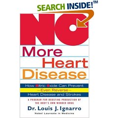
More reading about l-arginine and nitric oxide is available through Nobel laureate, Dr. Louis Ignarro's book, NO More Heart Disease : How Nitric Oxide Can Prevent--Even Reverse--Heart Disease and Stroke, available at Amazon.com ( http://www.amazon.com/gp/product/0312335814/104-1247258-6443909?v=glance&n=283155).


Will l-arginine truly reverse heart disease on its own? No, I don't believe so. Contrary to Dr. Ignarro's extravagant claims, I find l-arginine a facilitator of plaque regression, i.e, it helps other strategies achieve regression, but it does not achieve regression or reversal by itself. (Note that Dr. Ignarro is a lab researcher who studies rats and has never treated a human being.)

But l-arginine may have special application in the person with lp(a), particularly if hypertension is part of the syndrome.


Note: As always, please note that I talk frankly about l-arginine and other supplements and medications but have no hidden agenda: I am not selling anything, nor am I affiliated with any source/website/store etc. that sells these products. If I advocate something, I do so because I truly believe it, not because I'm trying to sell something. I make this point because so much nonsense is propagated in the media because of profit-motive. That's not true here.

Dr. Ornish: Get with the program!


In the era up until the 1980s, most Americans indulged in excessive quantities of saturated fats: fried chickem, spare ribs, French fries, gravy, bacon, Crisco, butter, etc.

Along came people like Nathan Pritikin and Dr. Dean Ornish, both of whom were vocal advocates of a low-fat nutritional approach. In their programs, fat composed no more than 10% of calories. This represented a dramatic improvement--at the time.


In 2006, a low-fat diet is a perversion of health. It means over-reliance on breads, breakfast cereals, pasta, crackers, cookies, pretzels, etc., the foods that pack supermarket shelves and that now constitute 70-80% of most Americans' diet.

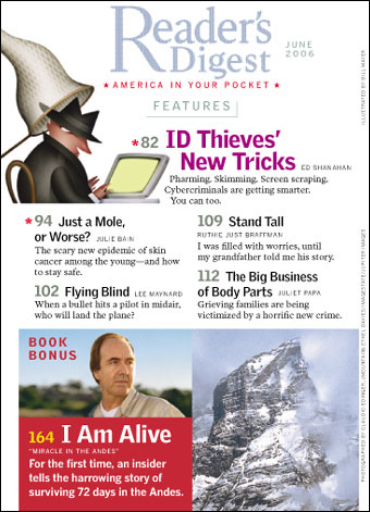
Dr. Ornish still carries great name recognition. As a result, his outdated concepts still gain media attention. The June, 2006 issue of Reader's Digest, in their RDHealth column, carried an interview with Dr. Ornish in which he reiterates his fat-phobia.

However, on this occasion he takes a different tack. This time he rails against the "dangers" of fish oil and omega-3 fatty acids. "I've recently learned that omega-3s are a double-edged sword...In some cases, omega-3s could be fatal."

He goes on to say that, while he believes that fish oil may prevent heart attacks, it has fatal effect if you already have heart disease.

Does this make sense to you?

He's basing his views on a single, obscure study published in 2003 conducted in rural England that showed an increase in death and heart attack on fish oil. Most authorities have not taken these findings seriously, since they are wildly contrary to all other observations and because the study had some design flaws.

Despite the fact that this isolated study runs counter to all other, better-conducted studies seems not to matter to Dr. Ornish.

Clinging to the low-fat concept is like hoping 8-track tapes will make a comeback. It's not going to happen. We enjoyed the benefits while they lasted, appropriate for the era. But now, they're woefully outdated.

The overwhelming evidence is that fish oil provides tremendous benefits with little or no downside. In the Track Your Plaque program, fish oil remains a crucial supplement to gain control over your coronary plaque and stop or reduce your heart scan score. Ignore the doomsday preachings of Dr. Ornish.

(Watch for an article I wrote updating the benefits of fish oil for Life Extension magazine.)

The cholesterol fallacy

Evan spotted the kiosk set up in the middle of the local mall. "Free cholesterol screenings. Know your heart health!" the sign declared.

It was a free cholesterol screening being offered by a local hospital.

The friendly nurse behind the kiosk had Evan fill out a form, then pricked his finger. Five minutes later, she reported to him with a smile, "Sir, your cholesterol is 177--your heart's fine! We get concerned when cholesterol is over 200. So you're in a safe range."

What the nurse failed to recognize is that Evan's HDL was 30 mg, a low value that actually places him at high risk for heart disease. Low HDL also signifies high likelihood of the small LDL particle pattern, a marked predisposition towards pre-diabetes and diabetes, a probable over-reliance on processed carbohydrates in his diet, a dramatically increased probability of hidden inflammation (e.g., elevated C-reactive protein), increased tendency for high blood pressure. . .

In other words, Evan's "favorable" total cholesterol is, in truth, nonsense. It's misleading, falsely reassuring, and provided none of the insight that a real effort might have yielded. Like hippies, tie-dye, other relics of the 1960s, total cholesterol needs to be put to rest. It has served many people poorly and been responsible for countless deaths.

When you see a kiosk or other service like this, even if it's free, run the other way.

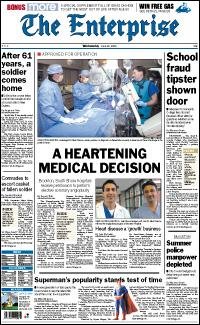
"Heart disease a growth business"


So announced a Boston newspaper recently, featuring a story about new heart program at a local hospital.

They were announcing how a hospital had entered the cardiovasculare procedure game and how it would boost their bottom line. The article discussed how the hospital administration was anticipating "a surge in patients from the baby boom generation."

To justify this new program, the article quoted an administrator from another hospital: "Cardiovascular issues is [sic] the number one cause people sought treatment at our hospital."

The hospital featured in the story had spent $13.5 million dollars to develop their program.

Do you think they'll make it back?

You bet they will--many times over. Hospitals are businesses, complete with a bottom line, an expectation of profit and an eye towards growth.

The hospitals in the city where I live (Milwaukee, Wisconsin) are, as in Boston and elsewhere, very aggressive--expanding into new territories, hiring new "salesmen" (physicians), all to capture more marketshare and produce more "product" (your coronary angioplasty, stent, bypass surgery, defibrillator, etc.).

The equation for hospital profits is tried and true. Ignore your heart disese risk and you can help your local hospital grow its business. Neglect to get your heart scan and you can help your hospital pay down its debt. Get a heart scan, then do nothing about it, and you may even justify a pay raise for the hospital administrators for record revenue growth and profit.

Hospitals are a growth business because of the failure of most people and their doctors to 1) identify hidden coronary disease (CT heart scan to obtain your heart scan score), then 2) seize control over it (the Track Your Plaque program or, at least, your doctor's guidance along with your efforts at prevention).

Unless you do so, you are highly likely to help your hospital boost its annual goal for procedures.

The myth of small LDL

Annie's doctor was puzzled.

Despite an HDL cholesterol of 76 mg (spectacular!) and LDL of 82 mg, her CT heart scan showed a score of 135. At age 51, this placed her in the 90th percentile.

Not as bad, perhaps, as her Dad might have had, since he died at age 54 of a heart attack.

So we submitted blood for lipoprotein testing. Surprise! over 90% of all her LDL particles were small. (By NMR, they're called "small". By gel electropheresis, or the Berkeley Lab test, or VAP (Atherotech) technique, they're called "HDL3".)

What gives? Traditional teaching in the lipid world is that if HDL equals or exceeds 40 mg/dl, then small LDL will simply not be present.

Well, as you can see from Annie's experience, this is plain wrong. Yes, there is a graded, population-based effect--the lower your HDL, the greater the likelihood of small LDL. But small LDL is remarkably persistent and prevalent--regardless of your HDL.

We've seen small LDL even with HDLs in the 90's! I call small LDL the "cockroach" of lipids. If you think you have it, you probably do. Getting rid of small LDL requires a specific bug killer. (Track Your Plaque Members: Read Dr. Tara Dall's interview on small LDL.)

Don't let anybody blow off your request for lipoprotein testing just because your HDL is high. That's just not acceptable. Loads can be wrong even with a favorable HDL.

My stress test was normal. I don't need a heart scan!

Katy had undergone a stress test while being seen in an emergency room, where she'd gone one weekend because of a dull pain on the right side of her chest. After her stress test proved normal, she was diagnosed (I believe correctly) with esophageal reflux, or regurgitation of stomach acid up the esophagus. She was prescrbed an acid-suppressing medication with complete relief.

But Katy also had coronary plaque. Three years ago, her CT heart scan score was 157. She'd made efforts to correct the multiple causes, though she still struggled with keeping weight down to gain full control over her small LDL particle pattern.

I felt it was time for a reassessment: another heart scan. After three years, without any preventive efforts, Katy's score would be expected to have reached 345! (That's 30% per year plaque growth.) It's a good idea to get feedback on just how much slowing you've accomplished.

But Katy declared, "But I didn't think another heart scan was necessary. My stress test was normal!"

What Katy was struggling to understand was that even at the time of her first scan, a stress test would have been normal. Plaque can be present with a normal stress test.

Plaque can even show explosive growth all while stress tests remain normal. Just ask former President, Bill Clinton, how much he should have relied on stress tests. (Mr. Clinton underwent annual stress nuclear tests. All were normal and he had no symptoms--all the way up 'til the time he needed urgent bypass surgery!)

Of course, at some point even a crude stress test will reveal abnormal results. But that's years into your disease and a lot closer to needing procedures and experiencing heart attack.

So, yes, Katy would benefit from another heart scan despite her normal stress test.

The message: Don't rely on stress tests to gauge whether or not plaque has grown, stabilized, or reversed. Stress tests can be used to gauge the safety of exercise, blood pressure response, and the potential for abnormal heart rhythms. Stress tests can be used as a method to determine whether blood flow in your coronary arteries is normal through an area with plaque.

But a stress test cannot be used to gauge whether plaque has grown. It's as simple as that. Gauging plaque growth requires a heart scan.

Patient-napping: Yet another reason to stay clear of hospitals!

When I started practicing medicine around 20 years ago, it was common practice to alert a physician when their patient was seen in an emergency room.

If John Smith, for example, went to the emergency room with chest pain, the physician who had an established relationship with the patient--knew their history, had managed their health and illnesses, etc.--was notified, even if the hospital ER had no relationship with the physician. It was not uncommon for the patient to then be transferred to the hospital where their own doctor practiced.

Though cumbersome at times, it preserved the relationship of the patient with their doctor.

Over the past few years, this practice has crumbled. Nowadays, hospitals and their employed physicians (and other unscrupulous physicians acting in the name of profit) "fail" to notify the physician with an established relationship.

Guess what happens? The patient all too often ends up being put through the gamut of testing and procedures.

Why? For hospital profit, of course. If failure to notify a doctor who's had a 10-year long relationship with the patient is "overlooked" or, even more commonly, it's "unsafe" to transfer the patient because the patient is too "unstable" to be transferred, then this patient becomes ripe for picking--heart catheterization, stents, bypass surgery, etc. Ten's, if not hundreds, of thousands of dollars can be reaped by this deception. I call it "patient-napping".

I see this at least several times every month. As hospitals are becoming increasingly competitive, and as they put pressure on their physicians to churn patients for revenues, you're going to see more and more of this.

As always, what is your protection from this expanding influence of hospitals and the doctors too meek to stand up to them? Education and information. Arm yourself with an understanding of what is accomplished in hospitals, when you truly need them, and when you don't.

Take it one step further. At least from a heart disease standpoint--the #1 profit-maker for hospitals--aim to 1)identify your coronary plaque, then 2) seize control over your coronary plaque and reduce your risk for heart attack and heart procedures as much as humanly possible. That's the goal of the Track Your Plaque program.

Don't believe the negative press on fish oil


A British Medical Journal study released in March, 2006 has prompted a media flurry of reports on the worthlessness of fish oil. (Hooper L, Thompson RL, Harrison RA et al. Risks and benefits of omega 3 fats for mortality, cardiovascular disease, and cancer: a systematic review. BMJ March,2006)

Don't believe it for a second.

First of all, the study was a re-analysis of the existing published scientific literature. It was not a new study. It included a wild conglomeration of different clinical observations, as the studies examining fish oil over the years have been extraordinarily heterogeneous--in populations examined, omega-3 supplement (e.g., fish vs. capsule), period of observation, endpoints measured.

The results were skewed by inclusion of a moderate-sized British study by Burr et al in men with angina. In this study, no benefit was demonstrated and, in fact, a negative effect--more heart attack and death--was observed with fish oil. This was not news, since the study was published in 2003. It's results have been a mystery to everyone, since its unexpected negative result for fish oil was so starkly different from virtually every other study that preceded it (suggesting a study flaw or statistical fluke).

Nonetheless, the Burr study served to throw off the overall analysis. It diluted the dramatic and persuasive outcome of the GISSI-Prevenzione Study of 11,000 people in which a 28% reduction in heart attack and 45% reduction in cardiovascular death was observed. Note that the substantial numbers of the GISSI make the study's outcome nearly unassailable.

Another important fact: fish oil is among the most powerful tools available to correct elevated triglycerides. Drops of 50% are common. Recall that triglycerides are a necessary ingredient to create the nasty LDL, as well as VLDL, Intermediate-density lipoprotein, and an undesirable shift from large to ineffective small HDL. Reducing triglycerides is therefore crucial for your plaque control program.

This re-analysis serves to prove nothing. Such analyses can only pose questions for further study in a real study like GISSI: a randomized (random participant assignment), controlled (treatment vs. placebo or other treatment) study.

The weight of evidence remains heavily in favor of fish oil, not only as helpful, but fabulously beneficial, particularly for anyone aiming to reduce coronary plaque.
Where do you find fructose?

Apple, 1 medium: Fructose 10.74 g


Honey: Fructose 17.19 grams per 2 tablespoons


Barbecue Sauce: HFCS number 1 ingredient
Ingredients: High Fructose Corn Syrup, Vinegar, Concentrated Tomato Juice (Water, Tomato Paste), Water, Modified Food Starch, Salt, Honey, Contains Less Than 2% of Molasses, Natural Flavor, Paprika, Spice, Mustard Flour, Guar Gum, Red 40.


A1 Steak Sauce: HFCS number 2 ingredient
Ingredients: Tomato puree (water, tomato paste), high fructose corn syrup, vinegar, salt, water dried onions, contains less than 2% of black pepper, modified food starch, citric acid, dried parsley, dried garlic, xanthan gum, caramel color, potassium sorbate and calcium disodium EDTA as preservatives, molasses, corn syrup, sugar, spices, tamarind, natural flavor

Do heart scans cause cancer?

Another in a series of data extrapolations that attempt to predict long-term cancer risk from medical radiation exposure was published in the July 13, 2009 Archives of Internal Medicine, viewable here.

Over the years, I've fussed about the radiation dose used by some centers for CT heart scans. (Note: I'm talking about CT heart scans, not CT coronary angiograms, an entirely different test with different radiation exposure.) In the "old" days, when electron-beam devices (EBT) were the best on the block, the old single-slice CT scanners (the predecessor of the current 64-slice MDCT scanners) exposed patients to ungodly quantities of radiation, while the EBT devices required very small quantities (0.5 mSv or about the equivalent of 4 standard chest x-rays or one mammogram).

But CT technology has advanced considerably. While EBT has been phased out (although it was an exceptional technology, GE acquired the small California manufacturer, then promptly scrapped the operation; you can guess why), multi-detector CT (MDCT) technology has improved in speed, image quality, and radiation exposure.

While it has improved, radiation exposure still remains an issue. The authors of the study applied the scanning protocols used at three hospitals and those in several CT heart scan studies, then calculated radiation exposure. They found a more than ten-fold range of exposure, from 0.8 mSv to 10.5 mSv. (All scanners were MDCT, none EBT.)

That's precisely what I've been worrying about: In the rapid rush to develop new devices, radiation exposure has often been a neglected issue. While some scan centers do an excellent job and take steps to minimize exposure, others barely lift a finger and consequently expose their patients to unnecessary radiation.

However, it's not as bad as it sounds. For one, the study included 16-slice MDCT scanners, a scanner type that I warned people to not use because of radiation. On the current most popular 64-slice devices, much lower radiation exposure is possible, on the order of 0.8-1.2 mSv routinely--if the center takes the effort.

This study, while eye-opening, will achieve some good: CT heart scans are here to stay. But the day-to-day practice of heart scanning should be:

1) standardized
2) conducted with radiation exposure as low as possible, preferably <0.8 mSv


To read more about this issue, below I've reprinted a 2007 full Track Your Plaque Special Report, CT Heart Scans and Radiation: The Real Story.


CT heart scans and radiation: The real story

“My personal opinion is that many patients today who are receiving multiple CT scans may well be getting at least comparable doses to subjects that have now developed malignancies from x-ray radiation received in the 1930s and '40s. And, similar to those days when the doses were unknown, the dose that patients receive today over a course of years of multiple CT scans is also completely unknown . . .

“I recommend that all healthcare providers become familiar with the concept that 1 in 1000 CT studies of the chest, abdomen, or pelvis may result in cancer.”


Richard C. Semelka, MD
Professor and Vice Chairman, Department of Radiology
University of North Carolina–Chapel Hill


Is this just hype to generate headlines? Or is the truth buried in the enormous marketing clout of the medical device industry, among which the imaging device manufacturers reign supreme?

It’s been over 110 years since radiation was first used for medical imaging. Over those years, it has had its share of misadventures.

In the 1930s and 1940s, before the dangers of radiation were recognized, shoe shoppers had shoes fitted using an x-ray device of the foot to assess fit. High doses of radiation were used to shrink enlarged tonsils and extinguish overactive thyroid glands. Attitudes towards radiation were so lax that doctors commonly permitted themselves to be exposed without protection day after day, year after year, until an unexpected rise in blood cancers like leukemia was observed. As recently as the 1970s and 1980s, cancers like Hodgkins’ disease were treated with high doses of radiation, also leading to radiation-induced diseases decades later.

Not all radiation is bad. Radiation can also be used as a therapeutic tool and even today remains a useful and reasonably effective method to reduce the size, sometimes eliminate, certain types of cancer. Forty percent of people with cancer now receive some form of radiation as part of their treatment (Ron E 2003).


Just how much does medical radiation add to our exposure?

Estimates vary, but most experts estimate that medical imaging provides approximately 15% of total lifetime exposure. In other words, radiation exposure from medical imaging is simply a small portion of total exposure that develops over the years of life. Exposure can be much higher, however, in a specific individual who undergoes repeated radiation imaging or treatment of one sort or another.

For all of us, exposure to medical radiation is part of lifetime exposure from multiple sources, added to the radiation we receive from the world around us. Just by living on earth, we are exposed to radiation from space and naturally-occurring radioactive compounds, and receive somewhere around 3.0 mSv per year (U.S. Nuclear Regulatory Commission). (Doses for radiation exposure are commonly expressed in milliSieverts, mSv, a measure that reflects whole-body radiation exposure.) People living in high-altitude locales like Colorado get exposed to an additional 30–50% ambient radiation (1.0–1.5 mSv more per year).

Much of the information on radiation exposure comes from studies like the Life Span Study that, since 1961, has tracked 120,000 Japanese exposed to radiation from the atomic bombs dropped in 1945 (Preston DL et al 2003). Although regarded as a high-dose exposure study for obvious reasons, there are actually thousands of people in this study who were exposed to lesser quantities of radiation (because of distance from the bomb sites) who still display a “dose-response” increased risk for cancer many years later in life. Radiation exposures of as little as 5–20 mSv showed a slight increase in lifetime risk.

Occupational and excessive medical exposure to radiation also provides a “laboratory” to examine radiation risk. Miners exposed to radon gas; patients exposed to the imaging agent, Thorotrast, containing radioactive isotope thorium dioxide and used as an x-ray contrast agent in the 1930s and 1940s and possesses the curious property of lingering in the body for over 30 years after administration; radium injections administered between 1945 and 1955 to treat diseases like ankylosing spondylitis and tuberculosis, all provide researchers an opportunity to study the long-term effects of various types of radiation exposure over many years (Harrison JD et al 2003).

The excess exposure of workers and several hundred thousand nearby residents to the Mayak nuclear plant in Russia has also revealed a “dose-response” relationship, with increasing exposure leading to more cancers, including leukemia and solid cancers of the bone, liver, and lung (Shilnikova NS et al 2003). Nuclear waste released into the Techa river between 1948 and 1956 contaminated drinking water used by over 100,000 Russians. A plant explosion in 1957 also released an excess of radiation into the atmosphere, yielding exposure via inhalation. Some sources estimate that at least 272,000 people have been affected by radiation from the Mayak plant. This unfortunate situation has, however, yielded plenty of data on radiation exposure and its long-term effects.

It’s also been known for several decades that people who receive therapeutic radiation for treatment of cancer, even with the reduced doses now employed, are subject to increased risk of a second cancer consequent to the radiation treatment.

From experiences like this, radiation experts estimate that an exposure of 10 mSv increases a population’s risk for cancer by 1 in 1000 (Semelka RC et al 2007).

This question was recently thrust into the spotlight with publication of a study from Columbia University in New York suggesting that a 20-year old woman would be exposed to a lifetime risk of cancer as high as 1 in 143 consequent to the radiation received during a CT coronary angiogram. (Important note: This was estimated risk from a CT coronary angiogram, not a simple heart scan that we advocate for the Track Your Plaque program.) The risk at the low end of the spectrum would be in an 80-year old man (because of the shorter period of time to develop cancer), with a risk of 1 in 5017. If “gating” to the EKG is added (which many scan centers do indeed perform nowadays), risk for a 60-year old woman is estimated at 1 in 715; risk for a 60-year old male, 1 in 1911 (Einstein AJ et al 2007). This study generated some criticism, since it did not directly involve human subjects, but used “phantoms” or x-ray dummies to simulate x-ray exposure. Nonetheless, the point was made: CT coronary angiograms in current practice do indeed expose the patient to substantial quantities of radiation, sufficient to pose a lifetime risk of cancer.


The media frenzy

The NY Times ran an article called With Rise in Radiation Exposure, Experts Urge Caution on Tests in which they stated:

"According to a new study, the per-capita dose of ionizing radiation from clinical imaging exams in the United States increased almost 600 percent from 1980 to 2006. In the past, natural background radiation was the leading source of human exposure; that has been displaced by diagnostic imaging procedures, the authors said."

“This is an absolutely sentinel event, a wake-up call,” said Dr. Fred A. Mettler Jr., principal investigator for the study, by the National Council on Radiation Protection. “Medical exposure now dwarfs that of all other sources.”

Radiation is a widely used imaging tool in medicine. Although CT scans of the brain, bones, chest, abdomen, and pelvis account for only 5% of all medical radiation procedures, they are responsible for nearly 50% of medical radiation used. It’s been known for years that increasing radiation exposure increases cancer risk over many years, but the boom of newer, faster devices that provide more detailed images has opened the floodgates to expanded use of CT scanners.

But before we join in the hysteria, let's first take a look at exposure measured for different sorts of tests:


Typical effective radiation dose values for common tests

Computed Tomography

Head CT 1 – 2 mSv
Pelvis CT 3 – 4 mSv
Chest CT 5 – 7 mSv
Abdomen CT 5 – 7 mSv
Abdomen/pelvis CT 8 – 11 mSv
Coronary CT angiography 5 – 12 mSv


Non-CT

Hand radiograph Less than 0.1 mSv
Chest radiograph Less than 0.1 mSv
Mammogram 0.3 – 0.6 mSv
Barium enema exam 3 – 6 mSv
Coronary angiogram 5 – 10 mSv
Sestamibi myocardial perfusion (per injection) 6 – 9 mSv
Thallium myocardial perfusion (per injection) 26 – 35 mSv

Source: Cynthia H. McCullough, Ph.D., Mayo Clinic, Rochester, MN


A plain, everyday chest x-ray, providing less than 0.1 mSv exposure, provides about the same quantity of radiation exposure as flying in an airplane for four hours, or the same amount of radiation from exposure to our surroundings for 11–12 days. Similar exposure arises from dental x-rays.

If you have a heart scan on an EBT device, then your exposure is 0.5-0.6 mSv, roughly the same as a mammogram or several standard chest x-rays.

With a heart scan on a 16- or 64-slice multidetector device, exposure is ideally around 1.0-2.0 mSv, about the same as 2-3 mammograms, though dose can vary with this technology depending on how it is performed (gated to the EKG, device settings, etc.)

CT coronary angiography presents a different story. This is where radiation really escalates and puts the radiation exposure issue in the spotlight. As Dr. Cynthia McCullough's chart shows above, the radiation exposure with CT coronary angiograms is 5-12 mSv, the equivalent of 100 or more chest x-rays or 20 mammograms. Now, that's a problem.

The exposure is about the same for a pelvic or abdominal CT. The problem is that some centers are using CT coronary angiograms as screening procedures and even advocating their use annually. This is where the alarm needs to be sounded. These tests, as wonderful as the information and image quality can be, are not screening tests. Just like a pelvic CT, they are diagnostic tests done for legitimate medical questions. They are not screening tests to be applied broadly and used year after year.

It’s also worth giving second thought to any full body scan you might be considering. These screening studies include scans of the chest, abdomen, and pelvis. These scans, performed for screening, expose the recipient to approximately 10 mSv of radiation (Radiological Society of North American, 2007). Debate continues on whether the radiation exposure is justified, given the generally asymptomatic people who generally undergo these tests.

Always be mindful of your radiation exposure, as the NY Times article rightly advises. However, don't be so frightened that you are kept from obtaining truly useful information from, for instance, a CT heart scan (not angiography) at a modest radiation cost.


Heart scans, CT coronary angiograms and the future

Unfortunately, practicing physicians and those involved in providing CT scans are generally unconcerned with radiation exposure. The majority, in fact, are entirely unaware of the dose of radiation required for most CT scan studies and unaware of the cancer risk involved. It is therefore up to the individual to insist on a discussion of the type of scanner being used, the radiation dose delivered (at least in general terms), the necessity of the test, alternative methods to obtain the same diagnostic information, all in the context of lifetime radiation exposure.

Our concerns about radiation exposure all boil down to concern over lifetime risk for cancer, a disease that strikes approximately 20% of all Americans. Many factors contribute to cancer risk, including obesity, excessive saturated fat intake, low fiber intake, lack of vitamin D, repeated sunburns, excessive alcohol use, smoking, exposure to pesticides and other organochemicals, asbestos and other industrial exposures, electromagnetic wave exposure, and genetics. Radiation is just one source of risk, though to some degree a controllable one.

Some people, on hearing this somewhat disturbing discussion, refuse to ever have another medical test requiring radiation. That’s the wrong attitude. It makes no more sense than wearing lead shielding on your body 24 hours a day to reduce exposure from the atmosphere. Taken in the larger context of life, radiation exposure is just one item on a list of potentially harmful factors.

It is, however, worth some effort to minimize radiation exposure over your lifetime, particularly before age 60, and by submitting to high-dose testing only when truly necessary, or when the potential benefits outweigh the risks. Thus, with heart scans and CT coronary angiography, some thought to the potential benefits of knowing your score or the information gained from the CT angiogram need to be considered before undergoing the test. Often the practical difficulty, of course, is that your risk for heart disease simply cannot be known until after the test.

In our view, in the vast majority of instances a simple CT heart scan can serve the simple but crucial role of quantifying risk for heart attack and atherosclerotic plaque. CT heart scans yield this information with less than a tenth of the radiation exposure of a CT coronary angiogram. In people without symptoms and a normal stress test, there is rarely a need for CT coronary angiography with present day levels of radiation exposure. Perhaps as technology advances and the radiation required to generate images is reduced, then we should reconsider.

Early experiences are suggesting that the newest 256-slice scanners, now being developed but not yet available, will cut the dose exposure of 64-slice CT angiograms in half (from 27.8 mSv to 14.1 mSv in a recent Japanese study). The 256-slice scanners will allow scanning that is faster over a larger area in a given period of time.

Thankfully, the scanner manufacturers are increasingly sensitive to the radiation issue and have been working on methods to reduce radiation exposure. However, it still remains substantial.


References:
Einstein AJ, Henzlova MJ, Rajagopalan S. Estimating risk of cancer associated with radiation exposure from 64-slice computed tomography coronary angiography. JAMA 2007 Jul 18;298(3):317–323.

Harrison JD, Muirhead CR. Quantitative comparisons of cancer induction in humans by internally deposited radionuclides and external radiation. Int J Radiat Biol 2003 Jan;79(1):1–13.

Hausleiter J, Meyer T, Hadamitzyky M et al. Radiation Dose Estimates From Cardiac Multislice Computed Tomography in Daily Practice: Impact of Different Scanning Protocols on Effective Dose Estimates. Circulation 2006;113:1305–1310.

Kalra MK, Maher MM, Toth TL, Hamberg LM, Blake MA, Shepard J, Saini S. Strategies for CT radiation dose optimization. Radiology 2004;230:619–628.

Mayo JR, Aldrich J, Müller NL. Radiation exposure at chest CT: A statement of the Fleischner Society. Radiology 2003; 228:15–21.

Mori S, Nishizawa K, Kondo C, Ohno M, Akahane K, Endo M. Effective doses in subjects undergoing computed tomography cardiac imaging with the 256-multislice CT scanner. Eur J Radiol 2007 Jul 10; [Epub ahead of print].

Preston DL, Pierce DA, Shimizu Y, Ron E, Mabuchi K. Dose response and temporal patterns of radiation-associated solid cancer risks. Health Phys 2003 Jul;85(1):43–46.

Ron E. Cancer risks from medical radiation. Health Phys 2003 Jul;85(1):47–59.

Shilnikova NS, Preston DL, Ron E et al. Cancer mortality risk among workers at the Mayak nuclear complex. Radiation Res 2003 Jun;159(6):787–798.

Semelka RC, Armao DM, Elias J Jr, Huda W. Imaging strategies to reduce the risk of radiation in CT studies, including selective substitution with MRI. J Magn Reson Imaging 2007 May;25(5):900–9090.


Copyright 2007, Track Your Plaque.

Goodbye, fructose

A carefully-conducted study by a collaborative research group at University of California-Berkeley has finally closed the lid on the fuss over fructose vs. glucose and its purported adverse effects.

The study is published in its entirety here.

Compared to glucose, fructose induced:

1) Four-fold greater intra-abdominal fat accumulation--3% increased intra-abdominal fat with glucose; 14.4% with fructose. (Intraabdominal fat is the variety that blocks insulin responses and causes diabetes and inflammation.)

2) 13.9% increase in LDL cholesterol but double the increase for Apoprotein B (an index of the number of LDL particles, similar to NMR LDL particle number).

3) 44.9% increase in small LDL, compared to 13.3% with glucose.

4) While glucose (curiously) reduced the net postprandial (after-eating) triglyceride response (area under the curve, AUC), fructose increased postprandial triglycerides 99.2%.


The authors propose that fructose specifically increases liver VLDL production, the lipoprotein particle that yields abnormal after-eating particles, increased LDL, and provides building blocks to manufacture small LDL particles. The authors also persuasively propose that fructose metabolism, unlike glucose, is not inhibited (via feedback loop) by energy intake, i.e., it's as if you are always starving.

Add to this the data that show that fructose increases uric acid (that causes gout and may act as a coronary risk factor), induces leptin resistance, causes metabolic syndrome (pre-diabetes), and increases appetite, and it is clear that fructose is yet another common food additive that, along with wheat, is likely a big part of the reason Americans are fat and diabetic.

Fructose is concentrated, of course, in high-fructose corn syrup, comprising anywhere from 42-90% of total weight. Fructose also composes 50% of sucrose (table sugar). Fructose also figures prominently in many fruits; among the worst culprits are raisins (30% fructose) and honey (41% fructose).

Also, beware of low-fat or non-fat salad dressings (rich with high-fructose corn syrup), ketchup, beer, fruit drinks, fruit juices, all of which are rich sources of this exceptionally fattening, metabolism-bypassing, LDL cholesterol/small LDL/ApoB increasing compound. Ironically, this means that many low-fat foods meant to reduce cholesterol actually increase it when they contain fructose in any form.

When you hear or say "fructose," run the other way, regardless of what the Corn Refiners Association says.

The statin-free life

Matt came to me because his doctor couldn't reduce his LDL cholesterol.

His doctor had prescribed Zocor (simvastatin), Lipitor, Crestor, even pravastatin, all of which resulted in incapacitating muscle aches and weakness within a week of starting. No surprise, Matt had a jaundiced view of statin drugs.

We started out by characterizing his lipoprotein patterns:

--LDL 155 mg/dl

--72% of LDL was small LDL, a moderately severe pattern. (This means that small LDL comprised 112 mg/dl of the total 155 mg/dl LDL; large LDL comprised 43 mg/dl--small LDL was the problem.)

--HDL 42 mg/dl --Triglycerides 133 mg/dl

--No lipoprotein(a)

Beyond lipoproteins, Matt proved severely deficient in vitamin D with a starting level of 18 ng/ml.

Matt's doctor had advised that he avoid salt, as his blood pressure had been borderline high. His thyroid assessment disclosed a TSH of 3.89 mIU/ml with thyroid hormones free T3 and free T4 in the lower half of the normal range.

I therefore asked Matt to:

--Eliminate wheat, cornstarch, and sugars to reduce small LDL
--Add iodine
--Supplement 6000 units of an oil-based vitamin D preparation
--Take fish oil to provide at least 1800 mg EPA + DHA per day
--Take Armour Thyroid 1 grain per day


Several months later on this program, Matt had a repeat basic lipid panel:

--LDL 82 mg/dl--a 47% reduction

--HDL 52 mg/dl a 24% increase

--Triglycerides 60 mg/dl--a 55% decrease

In addition, vitamin D was 66 ng/ml, TSH was <1.0 mIU/ml with free T3 and free T4 in the upper half of the "reference range." Matt also felt great.

While the numbers could be slightly better, Matt had made tremendous progress towards achieving perfect values.

There you have it: Marked correction of cholesterol values, no statin drugs involved.

Creatine: Not just for muscle heads

Even if you’re not interested in building big muscles like a bodybuilder, there are health benefits to increasing muscle mass: increased bone density, better balance, and fewer injuries. Greater muscle mass means higher metabolic rate, improved insulin responsiveness, lower blood sugar. The inevitable loss of muscle mass of aging can lead to frailty, an increasingly common situation for the elderly. Muscle loss be reversed, health improved as a result.

Since its introduction in 1994, creatine has exploded in popularity, particularly among bodybuilders and athletes interested in gaining muscle mass and strength. But creatine is not just for young weight lifters. If you are just interested in increasing muscle mass for its health benefits, then creatine is something to consider.

A study of creatine supplementation in men, average age 70 years, demonstrated that, when creatine was combined with strength training, it increased muscle mass 250% better than placebo (7.26 lb muscle vs 2.86 lb muscle), along with improved leg strength and endurance. The same group also demonstrated 3.2% increased bone density (measured using dual energy X-ray absorptiometry) after 12 weeks in participants taking creatine with strength training, while the control (no strength training, no creatine) group decreased by 1.0%.

Benefits are not confined to men. Similar results were observed in another study that included women (age 65 and older), with outcomes in females comparable to males. This is especially important for females, given the common development of osteopenia and osteoporosis in postmenopausal females.

Other studies have shown that benefits are maintained after stopping creatine supplementation.

The most popular form of creatine is the monohydrate, generally taken as a “loading” phase of 15-20 grams per day (generally split into 3-4 doses of 5 grams) for 5-7 days, followed by weeks to months of 2-5 grams per day.

An alternative form, polyethylene glycosylated creatine (PEG-creatine) provides similar effects at one-fourth to one-half the dose of creatine, i.e., 1.25-2.5 grams per day.

Despite previous concerns about kidney toxicity with prolonged use, another study showed that athletes taking creatine for up to 21 months have shown no adverse effects on kidney function, lipid (cholesterol) values, or other basic health measures.

Having healthy muscle mass doesn't make you bulge like a bodybuilder. With modest efforts at strength training, augmented with creatine supplementation, you have a wonderful tool to feel better, reduce injury, increase bone density, and combat abnormal insulin resistance, not to mention accelerate weight loss, since lean muscle mass consumes energy.

The ultimate “bioidentical” hormone

There has been a lot of debate over whether or not “bio-identical” hormones, i.e., hormones identical to the human form, are superior to non-human forms dispensed by the drug industry.

The FDA is currently taking steps to clamp down on availability of bioidentical hormones and their claims of superiority, despite a groundswell of grassroot support for them. The argument has pitted anti-aging practitioners and the public, as well as the likes of Oprah and Suzanne Somers, against Big Pharma and the FDA, the two forces trying to squash the bioidentical hormone movement.

Regardless of what heavy-handed approach the FDA takes, we already have access to hormones identical to the original human form. It requires no prescription and yields downstream hormones that the human body recognizes as human.

That "bioidentical" hormone is pregnenolone.

Pregnenolone is the first biochemical step in the conversion of dietary cholesterol (yes-cholesterol!) to numerous other hormones. Pregnenolone is the source of the hormones that lie at the center of the bioidentical hormone controversy: estrogens, progesterone, and testosterone. We therefore already have our own over-the-counter, non-prescription form of bioidentical hormones.

Supplemental pregnenolone increases estrogens (mildly), progesterone, and testosterone. Prenenonlone supplementation simply provide more of the basic substrate for hormone production. The increase in hormones is usually modest, not as vigorous as direct hormone replacement like, say, testosterone or progesterone topical creams. But pregnenolone can be useful when small to moderate increases are desired, such as for reduction of Lp(a). A theoretical downside is that pregnenonlone can also convert to cortisol, the adrenal gland hormone that regulates fluid and blood pressure. However, I've not seen any measurable increase in cortisol with low doses of pregnenonlone and limited data suggest that it does not. Pregnenolone also converts to the other adrenal gland hormone, DHEA; I call DHEA "the hormone of assertiveness," since some people who take too much pregnenolone (or direct DHEA) acquire excessive assertiveness.

The key to pregnenolone supplementation is to proceed gradually and begin with a small dose, e.g., 5 mg every morning. Hormonal assessment is best conducted periodically to assess the effects and to determine whether a dose adjustment is in order.

Roger's near-miss CT angiogram experience

Heart Scan Blog reader, Roger, described his near-miss experience with CT coronary angiograms.

Hoping to obtain just a simple CT heart scan, he was bullied to get a CT coronary angiogram instead. Roger held strong and just asked for the test that we all should be having, a CT heart scan.


I posted yesterday that I was about to have my first CT heart scan...well, it was an interesting experience for reasons I coudn't possibly have anticipated. Dr. Davis has commented in the past on the confusion in the media about the difference between a CT calcium score scan, and a CT angiography, the latter requiring a far higher dose of radiation. I assumed this was a source of confusion only among patients and lay folks, but, lo and behold, I discovered today that doctors--or at least their helpers--can be just as confused.

Here's my story:

After checking in, I asked the receptionist to see if she had any information on whether my medical insurance was covering the scan. She called someone, and I heard her say over the phone, "He's here for a CT angiogram." At that point my ears perked up. I explained I wasn't here for a CT angiogram, only a regular CT scan. "Well, do you want to call your doctor and talk about this?" she asked. No, I said, I would like to ask one of their folks to verify exactly what test my doctor had ordered. As luck would have it, the technician was walking by at that point. "Is this a CT angiogram?" the receptionist asked. "No, it's just a CT calcium score scan" was the reply. But apparently the technician had been unclear herself, and had called my doctor just to verify. In other words, the "default" procedure they were accustomed to doing at this august Houston vascular clinic was a CT angiogram.

In fact, my appointment was even listed on their calendar as a "CT angiogram." For all I know, my insurance will be billed for the same. Later, during the procedure, the technician acted surprised I wasn't doing the "full test." I explained I had minimal risk factors (actually only one, an HDL of 34 a couple of years ago, which has since been raised to 50 partly as a result of taking advice from this site), but that my doctor was progressive (he is an MD for the Houston Astros) and thought it was a good idea since there is heart disease in my immediate family. My doctor did indeed prescribe only a CT calcium score scan, but it seems to have been an order that this clinic, at least, wasn't all that used to seeing.

So, I guess the message is: we have a lot of educating to do. Had I not been a faithful reader of these pages, I certainly wouldn't have known what kind of test I was about to get, or what questions to ask!

As for the heart scan itself, a piece of cake. If you can hold your breath, you can take this test. Just be sure it is the right one!


Why the "push" towards CT coronary angiograms and not "just" a CT heart scan? Well, I know it's shocking but it's . . . money!

CT coronary angiograms yield around $1800-$4000 per test. CT heart scans yield somewhere around $200. Though the scan center support staff might not care too much about the money themselves, their administrators likely make the cost distinctions clear to them.

Another reason: Most scan center staff, ironically, don't understand what a heart scan means, nor do they understand how it might serve to launch a program of prevention. They do understand that severe blockage by CT angiogram "needs" to be stented or bypassed. So they push patients towards things they understand.

Nobody makes money from CT heart scans, just as nobody makes money from a mammogram. Heart scans also don't lead to heroic, "lifesaving" procedures. They just lead to this sleepy, unexciting, inexpensive thing called prevention.

The Myth of Prevention: Letter to the Wall Street Journal


The June 20-21, 2009 Wall Street Journal Weekend Journal featured a provocative front page article written by physician, Dr. Abraham Verghese:

The Myth of Prevention

While eloquently written, I took issue with a few crucial points. Here is the letter I sent to the Editor at Wall Street Journal:


Dear Wall Street Journal Editor,

Re: Dr. Abraham Verghese’s article, The Myth of Prevention in the June 20-21, 2009 Weekend Journal.


I believe a more suitable title for Dr. Verghese’s article would be: “The Myth of What Passes as Prevention.”

As a practicing cardiologist, I, too, have witnessed firsthand the systemic “corruption” described by Dr. Verghese, the doing things “to” people rather than “for” them. Heart care, in particular, is rife with this form of profit-driven health delivery.

There is a fundamental flaw in Dr. Verghese’s otherwise admirable analysis: He assumes that what is called “prevention” in mainstream medicine is truly preventive.

Dr. Verghese makes issue of the apparent minor differences between preventing a condition and just allowing a condition to run its course. Prostate cancer screening is one example: Men subjected to repeated screenings have little length-of-life advantage over men who just allow their prostate to suffer the expected course of disease.

What if, instead, “prevention” as practiced today is nothing more than a solution that has been adopted in mainstream practice to suit yet another doing “to” strategy than doing “for”? In the prostate cancer example, PSA and prostate exam screenings often serve as little more than a means of harvesting procedures for the local urologist.

That’s not prevention. It is a prototypical example of “prevention” being subverted into the cause of revenue-generating procedures.

I submit that Dr. Verghese has fallen victim to the very same system he criticizes. His views have unwittingly been corrupted by the corrupt profit-driven system he describes.

What if, instead, prevention were just that: prevention or elimination of the condition. What if “prevention” of prostate cancer eliminated prostate cancer? What if heart disease “prevention” prevented all heart disease? What if this all proceeded without regard for profit or revenue-generating procedures, but just on results?

Dr. Verghese specifically targets heart scans or coronary calcium scoring, a test he likens to “miracle glow-in-the-dark minnow lures,” calling them “moneymakers.” Yes, when subverted into a corrupt algorithm of stress test, heart catheterization, stent, or bypass, heart scans are indeed a test used wrongly to “prevent” heart disease.

But what if the risk insights provided by heart scans prompt the start of a benign yet effective “prevention” program that inexpensively, safely, and assuredly prevents--in the true sense of the word--or eliminates heart disease? Then I believe the differences in mortality, quality of life, and costs would be substantial. Such strategies exist, yet do not necessarily include prescription drugs and certainly do not include the aftermath of heart catheterization and bypass surgery. Yet such programs fail to seize the limelight of media attention with no new high-tech lifesaving headline nor a big marketing budget to broadcast its message.

The problem in medicine is not prevention and its failure to yield cost- and life-saving results. It is the pervasively profit-driven mindset that keeps true preventive strategies from entering mainstream conversation. It is a repeat of Dr. Ignaz Semmelweis’ late 19th-century pleads for physicians to wash their hands before delivering babies to reduce puerperal sepsis, ignominious advice that earned him life and death in an asylum. We are essentially continuing to deliver children with unwashed hands because there is no revenue-generating procedure to clean them.

No, Dr. Verghese, the economic and medical failings of preventive strategies are not at fault. The failure of the medical system, in which everyone is bent on seizing a piece of the financial action for himself, has resulted in the failure to support the propagation of true preventive strategies that could genuinely save money and lives.

President Obama’s goal of cultivating preventive practices in medicine can work, but only if the profit-motive for “prevention” does not serve as the primary determinant of practice. Results-driven practices that are applied without regard to profit have the potential to yield the sorts of cost-saving and life-saving results that can reduce healthcare costs.


William Davis, MD
Milwaukee, Wisconsin
Medical Director, The Track Your Plaque Program (www.cureality.com)
Blog: http://heartscanblog.blogspot.com

A victory for SHAPE, CT heart scans, and doing what is RIGHT

The efforts of Texas House of Representatives Rep. Rene Oliveira and the SHAPE Guidelines committee have paid off: The Texas legislature passed a bill that requires health insurers to cover CT heart scans.

(NOTE: Don't make the same mistake that the media often makes and confuse CT heart scans with CT coronary angiography: two different tests, two different results, two different levels of radiation exposure. The difference is discussed here.)

Track Your Plaque previously reported the release of the SHAPE Guidelines, an ambitious effort to open CT heart scanning to people who would benefit from a simple screening test for coronary disease. Rep. Rene Oliveira initially introduced the bill in 2006, after having a heart scan uncovered extensive coronary plaque that resulted in coronary bypass surgery.

The bill requires that health-benefit providers cover the cost of CT heart scans (and carotid ultrasound) in men between the ages of 45-76, women 55-76, as well as anyone with diabetes or at "intermediate-risk" or higher for coronary disease by Framingham risk score.

The usual panel of cardiology knuckleheads stepped to the media podium, expressing their incredulity that something as "unvalidated" as heart scans could gain the backing of legislative mandate. Heartwire carried this comment:

"Contacted by heartwire, Dr Amit Khera (University of Texas Southwestern Medical Center, Dallas) confirmed there are still no comprehensive, adequately powered studies showing that these screening tests lead to better outcomes. In a phone interview, Khera said he has major concerns about how physicians will use these tests, particularly primary-care physicians. "I gave a talk last week to primary-care doctors, and there were probably 250 people in the room, and when I asked how many people had ordered a calcium scan, just one person raised a hand. . . . Most people don't even know what to do with the Framingham risk score, so they're going to follow an algorithm that they don't know how to follow to order a test result that they don't know what to do with."

It's the same criticisms hurled at heart scans over the years despite literally thousands of studies validating their application.

Studies have conclusively shown that:

--Coronary calcium scores generated by a CT heart scan outperform any other risk measure for coronary disease, including LDL cholesterol, c-reactive protein, total cholesterol, HDL cholesterol, blood pressure.
--Coronary calcium scores yield a graded, trackable index of coronary risk. Scores that increase correlate with increased risk of cardiovascular events; scores that remain unchanged correlate with much reduced risk.
--A coronary calcium score of zero--no detectable calcium--correlates with extremely low 5-year risk for cardiovascular events.
--Coronary calcium scores correlate with other measures of coronary disease. Heart scans correlate with coronary angiography, quantitative coronary angiography, carotid ultrasound (intimal-medial thickness and plaque severity), ankle-brachial index, and stress tests, including radionuclide (nuclear) perfusion imaging.

The reluctance of my colleagues to embrace heart scans stems from two issues, for the most part:

1) No study has yet been performed showing that knowing what the score is vs. not knowing what the score is changes prognosis. That's true. But it is also true of the great majority of practices in medicine. While many wrongs don't make a right, the miserable and widespread failure of other coronary risk measures, like LDL cholesterol or c-reactive protein, to readily and reliably detect hidden coronary disease creates a gaping void for improved efforts at early detection. If your LDL cholesterol is 140 mg/dl, do you or don't you have coronary disease? If your doctor's response is "Just take a statin drug anyway" you've been done a great disservice. (If and when this sort of study gets done, its huge cost--outcome studies have to be large and last many years--it will likely be a statin study. It is unlikely it will include such Track Your Plaque strategies that help reduce heart scan scores, like vitamin D and correction of small LDL particles.)

2) Fears over overuse of hospital procedures triggered by heart scans. This is a legitimate concern--if the information provided by a heart scan is misused. Heart scans should never--NEVER--lead directly to heart catheterization, stents, bypass surgery. Heart scans do not change the indications for performing revascularization (angioplasty, stents, bypass). Just because 20% of my cardiology colleagues are more concerned with profit rather than patient welfare does not invalidate the value of the test. Just because the mechanic at the local garage gouged you by replacing a carburetor for $800 when all you need was a new spark plug does not mean that we should outlaw all auto mechanics. Abuse is the fault of the abuser, not of the tool used to exercise the abuse.


All in all, while I am not a fan of legislating behavior in healthcare, the blatant and extreme ignorance of this simple tool for uncovering hidden heart disease makes the Texas action a huge success for heart disease prevention. I hope that this success will raise awareness, not just in Texas, but in other states and cities in which similar systemic neglect is the rule.

Remember: CT heart scans are tools for prevention, not to uncover "need" for procedures. They serve as a starting point to decide whether or not an intensive program of prevention is in order, and I don't mean statin vs. no statin.

Though not a multi-million dollar statin drug study, I have NEVER seen a heart attack or "need" for procedure in any person who has stopped progression or reduced their heart scan score. A small cohort from my practice was reported:

Effect of a Combined Therapeutic Approach of Intensive Lipid Management, Omega-3 Fatty Acid Supplementation, and Increased Serum 25 (OH) Vitamin D on Coronary Calcium Scores in Asymptomatic Adults.

Davis W, Rockway S, Kwasny M.

The impact of intensive lipid management, omega-3 fatty acid, and vitamin D3 supplementation on atherosclerotic plaque was assessed through serial computed tomography coronary calcium scoring (CCS). Low-density lipoprotein cholesterol reduction with statin therapy has not been shown to reduce or slow progression of serial CCS in several recent studies, casting doubt on the usefulness of this approach for tracking atherosclerotic progression. In an open-label study, 45 male and female subjects with CCS of >/= 50 without symptoms of heart disease were treated with statin therapy, niacin, and omega-3 fatty acid supplementation to achieve low-density lipoprotein cholesterol and triglycerides /=60 mg/dL; and vitamin D3 supplementation to achieve serum levels of >/=50 ng/mL 25(OH) vitamin D, in addition to diet advice. Lipid profiles of subjects were significantly changed as follows: total cholesterol -24%, low-density lipoprotein -41%; triglycerides -42%, high-density lipoprotein +19%, and mean serum 25(OH) vitamin D levels +83%. After a mean of 18 months, 20 subjects experienced decrease in CCS with mean change of -14.5% (range 0% to -64%); 22 subjects experienced no change or slow annual rate of CCS increase of +12% (range 1%-29%). Only 3 subjects experienced annual CCS progression exceeding 29% (44%-71%). Despite wide variation in response, substantial reduction of CCS was achieved in 44% of subjects and slowed plaque growth in 49% of the subjects applying a broad treatment program.

Sleep: A to Zzzzzzzzzz

Take a look at the results from the Heart Scan Blog's most recent reader poll (399 respondents):

How many hours do you sleep per night (on average)?


9 or more hours per night
15 (3.7%)

8-9 hours per night
72 (18%)

7-8 hours per night
152 (38.1%)

6-7 hours per night
111 (27.8%)

5-6 hours per night
38 (9.5%)

Less than 5 hours per night
11 (2.8%)


Like many issues in health, too much or too little of a good thing can present undesirable consequences.

Too much sleep: While psychologists and sleep researchers advise us that at least 9 hours are required to fully eliminate sleep "debt" and achieve optimal vigilance and mental performance, epidemiologic studies have shown increased mortality with this quantity of sleep.

Too little sleep: Getting less than 7 hours habituallly increases blood sugar, appetite, inflammatory measures, and encourages weight gain. Mortality is also increased, just as with sleeping too much. It is also associated with increased likelihood of a positive heart scan score.

7-8 hours per night from a health viewpoint is that Goldlilocks "just right" value: just enough to not erode mental performance substantially, but not so little that inflammatory, insulin-disrupting, and appetite-increasing effects develop.

Of our 399 respondents in the poll, 56.1% (38% + 18%) slept what appears to be an optimal amount for health. While only 3.7% slept too much (9 hours or more), the remaining 40.1% slept too little.

Our informal poll confirms what most of us observe in everyday life: The majority of people shortchange sleep in order to meet the demands of their high-pressure, squeeze-as-much-as-possible-into-every-day lives. But not paying off your sleep "debt" is like not paying the mortgage for a couple of months. You wouldn't expect your friendly neighborhood bank to say, "Oh, you forgot to pay your mortgage? Forget about it. Just pay next month's." Sure, fat chance. But if you don't pay off your sleep "debt," you will pay it back with health.